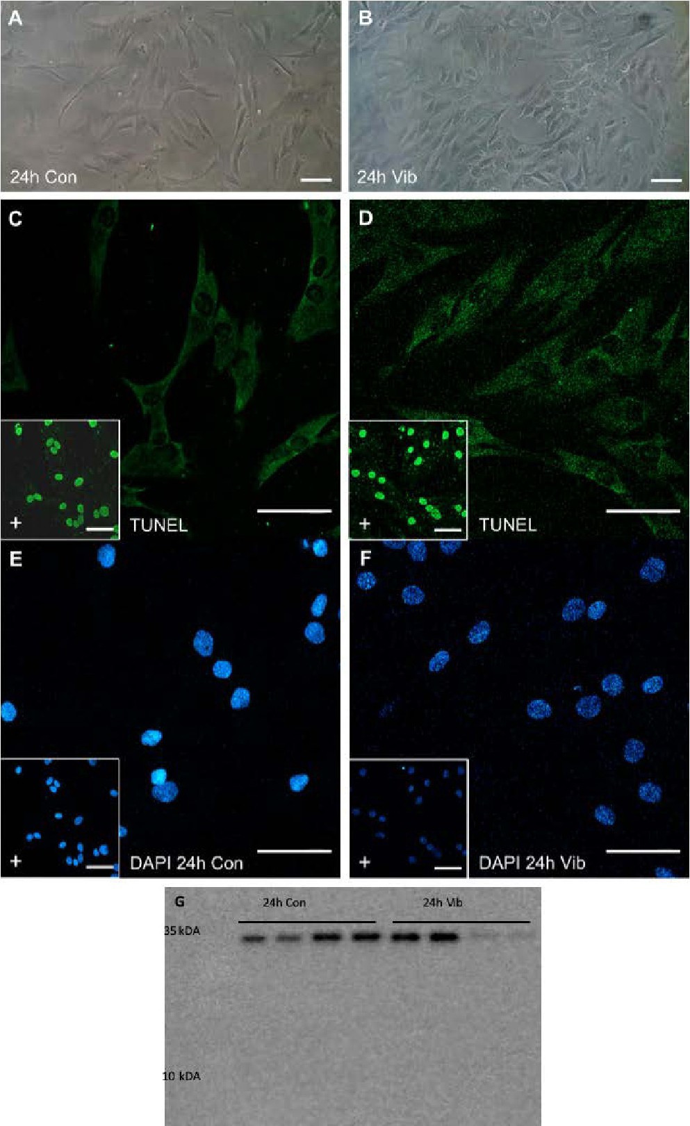
Fig. 1. Morphology of human primary chondrocytes in (A) static controls and (B) after 24h of VIB. TUNEL assay in comparison to DAPI counter staining (C, D) was used for apoptosis detection (green fluorescence). Positive controls (inserts) were initiated by DNase 1 prior to staining. DAPI staining (E, F) after fixation with PFA indicated no apoptotic bodies inside the nuclei or decomposition. (G) No cleaved caspase-3 (17 kDa) could be detected by Western blot. The uncleaved Cas-3 band visible at 32 kDa. Scale bars: 100 µm.